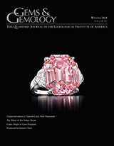
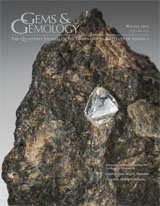
and pavilion (right) views of a chameleon diamond with visible trigons and tetragons. The trigons and tetragons are shown in more detail in figures 2 and 3. The diamond measures 1.28 cm in length. Photo by Adriana Robinson.")
Trigons and tetragons observed in a chameleon diamond indicate both octahedral and cubic growth.
Read More phosphorescence images of the table (left) and pavilion (right) in a 0.90 ct D-color natural diamond with very low boron showed localized areas of the 500 nm (greenish blue) and 660 nm (red) bands. Images by Evelina Goldort.")
A rare observation of spatially distinct red and greenish blue phosphorescence in a natural diamond.
Read More
A natural type IIa diamond displays a unique fluorescence pattern when exposed to deep-UV light.
Read More
The first report of an etch channel–like structure in a CVD laboratory-grown diamond submitted to GIA.
Read More fluorescence image showed blue coloration (due to the presence of dislocation bundles) that appeared nominally similar to DiamondView images of natural type IIa diamonds. Right: When the fluorescence was filtered by an orange long-pass filter that blocked wavelengths below 550 nm, the NV-related fluorescence and the striations indicative of CVD growth became apparent. Image by Sally Eaton-Magaña (right).")
A CVD-grown diamond submitted to GIA requires very careful examination to determine its origin.
Read More
The quality and size of this 4.04 ct CVD-grown diamond ring demonstrate the advancing technology in laboratory-grown diamonds.
Read More
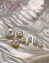
GIA’s Mumbai laboratory reports on the examination of an 80-year-old collection of pearls from the Arabian (Persian) Gulf.
Read More
A white button-shaped nacreous pearl displays rare flame-like structure.
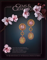
Read More set with colored gemstones and pearls measuring approximately 6.63 × 6.09 mm to 8.42 mm (left) and 5.38 mm to 8.02 × 7.61 mm (right). Photo by Gaurav Bera.")
Two Indian-style pearl nose rings are examined in the Mumbai laboratory.
Read More
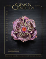
Fingerprints resulting from a flux-assisted heating process give a purple-red ruby a unique appearance.
Read Morepast gems & gemology issues

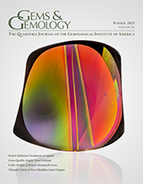
Fall 2023
Volume 59, Issue 3
Summer 2023
Volume 59, Issue 2

Spring 2023
Volume 59, Issue 1

Winter 2022
Volume 58, Issue 4

Fall 2022
Volume 58, Issue 3

Summer 2022
Volume 58, Issue 2

Spring 2022
Volume 58, Issue 1

Winter 2021
Volume 57, Issue 4
Fall 2021
Volume 57, Issue 3

Summer 2021
Volume 57, Issue 2
Spring 2021
Volume 57, Issue 1

Winter 2020
Volume 56, Issue 4

Fall 2020
Volume 56, Issue 3

Summer 2020
Volume 56, Issue 2

Spring 2020
Volume 56, Issue 1

Winter 2019
Volume 55, Issue 4

Fall 2019
Volume 55, Issue 3

Summer 2019
Volume 55, Issue 2

Spring 2019
Volume 55, Issue 1
Winter 2018
Volume 54, Issue 4

Fall 2018
Volume 54, Issue 3
Summer 2018
Volume 54, Issue 2

Spring 2018
Volume 54, Issue 1
Winter 2017
Volume 53, Issue 4

Fall 2017
Volume 53, Issue 3
Summer 2017
Volume 53, Issue 2

Spring 2017
Volume 53, Issue 1

Winter 2016
Volume 52, Issue 4

Fall 2016
Volume 52, Issue 3
Summer 2016
Volume 52, Issue 2

Spring 2016
Volume 52, Issue 1

Winter 2015
Volume 51, Issue 4

Fall 2015
Volume 51, Issue 3
Summer 2015
Volume 51, Issue 2

Spring 2015
Volume 51, Issue 1

Winter 2014
Volume 50, Issue 4

Fall 2014
Volume 50, Issue 3

Summer 2014
Volume 50, Issue 2

Spring 2014
Volume 50, Issue 1
Winter 2013
Volume 49, Issue 4

Fall 2013
Volume 49, Issue 3

Summer 2013
Volume 49, Issue 2

Spring 2013
Vol. 49, No. 1
Winter 2012
Volume 48, Issue 4

Fall 2012
Volume 48, Issue 3

Summer 2012
Volume 48, Issue 2

Spring 2012
Volume 48, Issue 1

Winter 2011
Volume 47, Issue 4

Fall 2011
Volume 47, Issue 3
Summer 2011
Volume 47, Issue 2

Spring 2011
Volume 47, Issue 1

Winter 2010
Volume 46, Issue 4

Fall 2010
Volume 46, Issue 3

Summer 2010
Volume 46, Issue 2
Spring 2010
Volume 46, Issue 1
Winter 2009
Volume 45, Issue 4
Fall 2009
Volume 45, Issue 3

Summer 2009
Volume 45, Issue 2

Spring 2009
Volume 45, Issue 1

Winter 2008
Volume 44, Issue 4

Fall 2008
Volume 44, Issue 3
Summer 2008
Volume 44, Issue 2

Spring 2008
Volume 44, Issue 1

Winter 2007
Volume 43, Issue 4

Fall 2007
Volume 43, Issue 3

Summer 2007
Volume 43, Issue 2

Spring 2007
Volume 43, Issue 1

Winter 2006
Volume 42, Issue 4

Fall 2006
Volume 42, Issue 3
Summer 2006
Volume 42, Issue 2

Spring 2006
Volume 42, Issue 1

Winter 2005
Volume 41, Issue 4
Fall 2005
Volume 41, Issue 3
Summer 2005
Volume 41, Issue 2

Spring 2005
Volume 41, Issue 1
Winter 2004
Volume 40, Issue 4

Fall 2004
Volume 40, Issue 3

Summer 2004
Volume 40, Issue 2

Spring 2004
Volume 40, Issue 1

Winter 2003
Volume 39, Issue 4

Fall 2003
Volume 39, Issue 3
Summer 2003
Volume 39, Issue 2
Spring 2003
Volume 39, Issue 1

Winter 2002
Volume 38, Issue 4
Fall 2002
Volume 38, Issue 3

Summer 2002
Volume 38, Issue 2

Spring 2002
Volume 38, Issue 1

Winter 2001
Volume 37, Issue 4

Fall 2001
Volume 37, Issue 3

Summer 2001
Volume 37, Issue 2
Spring 2001
Volume 37, Issue 1

Winter 2000
Volume 36, Issue 4
Fall 2000
Volume 36, Issue 3

Summer 2000
Volume 36, Issue 2
Spring 2000
Volume 36, Issue 1
Winter 1999
Volume 35, Issue 4

Fall 1999
Volume 35, Issue 3

Summer 1999
Volume 35, Issue 2

Spring 1999
Volume 35, Issue 1

Winter 1998
Volume 34, Issue 4
Fall 1998
Volume 34, Issue 3
Summer 1998
Volume 34, Issue 2
Spring 1998
Volume 34, Issue 1

Winter 1997
Volume 33, Issue 4
Fall 1997
Volume 33, Issue 3
Summer 1997
Volume 33, Issue 2
Spring 1997
Volume 33, Issue 1

Winter 1996
Volume 32, Issue 4

Fall 1996
Volume 32, Issue 3

Summer 1996
Volume 32, Issue 2
Spring 1996
Volume 32, Issue 1

Winter 1995
Volume 31, Issue 4
Fall 1995
Volume 31, Issue 3

Summer 1995
Volume 31, Issue 2

Spring 1995
Volume 31, Issue 1
Winter 1994
Volume 30, Issue 4
Fall 1994
Volume 30, Issue 3
Summer 1994
Volume 30, Issue 2

Spring 1994
Volume 30, Issue 1

Winter 1993
Volume 29, Issue 4

Fall 1993
Volume 29, Issue 3
Summer 1993
Volume 29, Issue 2

Spring 1993
Volume 29, Issue 1

Winter 1992
Volume 28, Issue 4

Fall 1992
Volume 28, Issue 3
Summer 1992
Volume 28, Issue 2
Spring 1992
Volume 28, Issue 1
Winter 1991
Volume 27, Issue 4
Fall 1991
Volume 27, Issue 3

Summer 1991
Volume 27, Issue 2
Spring 1991
Volume 27, Issue 1
Winter 1990
Volume 26, Issue 4

Fall 1990
Volume 26, Issue 3

Summer 1990
Volume 26, Issue 2

Spring 1990
Volume 26, Issue 1

Winter 1989
Volume 25, Issue 4
Fall 1989
Volume 25, Issue 3

Summer 1989
Volume 25, Issue 2
Spring 1989
Volume 25, Issue 1
Winter 1988
Volume 24, Issue 4

Fall 1988
Volume 24, Issue 3

Summer 1988
Volume 24, Issue 2

Spring 1988
Volume 24, Issue 1

Winter 1987
Volume 23, Issue 4
Fall 1987
Volume 23, Issue 3

Summer 1987
Volume 23, Issue 2

Spring 1987
Volume 23, Issue 1
Winter 1986
Volume 22, Issue 4
Fall 1986
Volume 22, Issue 3

Summer 1986
Volume 22, Issue 2

Spring 1986
Volume 22, Issue 1

Winter 1985
Volume 21, Issue 4

Fall 1985
Volume 21, Issue 3
Summer 1985
Volume 21, Issue 2

Spring 1985
Volume 21, Issue 1

Winter 1984
Volume 20, Issue 4
Fall 1984
Volume 20, Issue 3
Summer 1984
Volume 20, Issue 2

Spring 1984
Volume 20, Issue 1

Winter 1983
Volume 19, Issue 4

Fall 1983
Volume 19, Issue 3
Summer 1983
Volume 19, Issue 2
Spring 1983
Volume 19, Issue 1
Winter 1982
Volume 18, Issue 4
Fall 1982
Volume 18, Issue 3

Summer 1982
Volume 18, Issue 2

Spring 1982
Volume 18, Issue 1

Winter 1981
Volume 17, Issue 4
Fall 1981
Volume 17, Issue 3

Summer 1981
Volume 17, Issue 2
Spring 1981
Volume 17, Issue 1

Winter 1980
Volume 16, Issue 12

Fall 1980
Volume 16, Issue 11

Summer 1980
Volume 16, Issue 10
Spring 1980
Volume 16, Issue 9

Winter 1979
Volume 16, Issue 8
Fall 1979
Volume 16, Issue 7

Summer 1979
Volume 16, Issue 6

Spring 1979
Volume 16, Issue 5

Winter 1978
Volume 16, Issue 4

Fall 1978
Volume 16, Issue 3

Summer 1978
Volume 16, Issue 2

Spring 1978
Volume 16, Issue 1

Winter 1977
Volume 15, Issue 12

Fall 1977
Volume 15, Issue 11

Summer 1977
Volume 15, Issue 10

Spring 1977
Volume 15, Issue 9

Winter 1976
Volume 15, Issue 8

Fall 1976
Volume 15, Issue 7

Summer 1976
Volume 15, Issue 6
Spring 1976
Volume 15, Issue 5

Winter 1975
Volume 15, Issue 4

Fall 1975
Volume 15, Issue 3

Summer 1975
Volume 15, Issue 2

Spring 1975
Volume 15, Issue 1

Winter 1974
Volume 14, Issue 12
Fall 1974
Volume 14, Issue 11

Summer 1974
Volume 14, Issue 10
Spring 1974
Volume 14, Issue 9
Winter 1973
Volume 14, Issue 8

Fall 1973
Volume 14, Issue 7

Summer 1973
Volume 14, Issue 6

Spring 1973
Volume 14, Issue 5

Winter 1972
Volume 14, Issue 4

Fall 1972
Volume 14, Issue 3

Summer 1972
Volume 14, Issue 2

Spring 1972
Volume 14, Issue 1

Winter 1971
Volume 13, Issue 12

Fall 1971
Volume 13, Issue 11

Summer 1971
Volume 13, Issue 10
Spring 1971
Volume 13, Issue 9

Winter 1970
Volume 13, Issue 8

Fall 1970
Volume 13, Issue 7

Summer 1970
Volume 13, Issue 6
Spring 1970
Volume 13, Issue 5
Winter 1969
Volume 13, Issue 4

Fall 1969
Volume 13, Issue 3

Summer 1969
Volume 13, Issue 2

Spring 1969
Volume 13, Issue 1
Winter 1968
Volume 12, Issue 12
Fall 1968
Volume 12, Issue 11

Summer 1968
Volume 12, Issue 10
Spring 1968
Volume 12, Issue 9
Winter 1967
Volume 12, Issue 8

Fall 1967
Volume 12, Issue 7
Summer 1967
Volume 12, Issue 6

Spring 1967
Volume 12, Issue 5
Winter 1966
Volume 12, Issue 4

Fall 1966
Volume 12, Issue 3

Summer 1966
Volume 12, Issue 2
Spring 1966
Volume 12, Issue 1

Winter 1965
Volume 11, Issue 12

Fall 1965
Volume 11, Issue 11

Summer 1965
Volume 11, Issue 10
Spring 1965
Volume 11, Issue 9

Winter 1964
Volume 11, Issue 8

Fall 1964
Volume 11, Issue 7
Summer 1964
Volume 11, Issue 6

Spring 1964
Volume 11, Issue 5

Winter 1963
Volume 11, Issue 4
Fall 1963
Volume 11, Issue 3

Summer 1963
Volume 11, Issue 2

Spring 1963
Volume 11, Issue 1
Winter 1962
Volume 10, Issue 12
Fall 1962
Volume 10, Issue 11

Summer 1962
Volume 10, Issue 10
Spring 1962
Volume 10, Issue 9
Winter 1961
Volume 10, Issue 8

Fall 1961
Volume 10, Issue 7

Summer 1961
Volume 10, Issue 6
Spring 1961
Volume 10, Issue 5

Winter 1960
Volume 10, Issue 4

Fall 1960
Volume 10, Issue 3

Summer 1960
Volume 10, Issue 2
Spring 1960
Volume 10, Issue 1

Winter 1959
Volume 9, Issue 12
Fall 1959
Volume 9, Issue 11

Summer 1959
Volume 9, Issue 10

Spring 1959
Volume 9, Issue 9

Winter 1958
Volume 9, Issue 8

Fall 1958
Volume 9, Issue 7

Summer 1958
Volume 9, Issue 6
Spring 1958
Volume 9, Issue 5
Winter 1957
Volume 9, Issue 4

Fall 1957
Volume 9, Issue 3

Summer 1957
Volume 9, Issue 2
Spring 1957
Volume 9, Issue 1

Winter 1956
Volume 8, Issue 12

Fall 1956
Volume 8, Issue 11

Summer 1956
Volume 8, Issue 10

Spring 1956
Volume 8, Issue 9
Winter 1955
Volume 8, Issue 8
Fall 1955
Volume 8, Issue 7

Summer 1955
Volume 8, Issue 6
Spring 1955
Volume 8, Issue 5
Winter 1954
Volumt 8, Issue 4

Fall 1954
Volume 8, Issue 3
Summer 1954
Volume 8, Issue 2
Spring 1954
Volume 8, Issue 1
Winter 1953
Volume 7, Issue 12

Fall 1953
Volume 7, Issue 11

Summer 1953
Volume 7, Issue 10

Spring 1953
Volume 7, Issue 9

Winter 1952
Volume 7, Issue 8
Fall 1952
Volume 7, Issue 7

Summer 1952
Volume 7, Issue 6

Spring 1952
Volume 7, Issue 5
Winter 1951
Volume 7, Issue 4

Fall 1951
Volume 7, Issue 3

Summer 1951
Volumt 7, Issue 2

Spring 1951
Volume 7, Number 1

Winter 1950
Volume 6, Issue 12
Fall 1950
Volume 6, Issue 11

Summer 1950
Volume 6, Issue 10
Spring 1950
Volume 6, Issue 9
Winter 1949
Volume 6, Issue 6
Fall 1949
Volume 6, Issue 7

Summer 1949
Volume 6, Issue 6
Spring 1949
Volume 6, Issue 5
Winter 1948
Volume 6, Issue 4
Fall 1948
Volume 6, Issue 3

Summer 1948
Volume 6, Issue 2
Spring 1948
Volume 6, Issue 1
Winter 1947
Volume 5, Issue 12
Fall 1947
Volume 5, Issue 11
Summer 1947
Volume 5, Issue 10
Spring 1947
Volume 5, Issue 9

Winter 1946
Volume 5, Issue 8
Fall 1946
Volume 5, Issue 7
Summer 1946
Volume 5, Issue 6

Spring 1946
Volume 5, Issue 5
Winter 1945
Volume 5, Issue 4

Fall 1945
Volume 5, Issue 3
Summer 1945
Volume 5, Issue 2
Spring 1945
Volume 5, Issue 1

Winter 1944
Volume 4, Number 12
Fall 1944
Volume 4, Issue 11
Summer 1944
Volume 4, Issue 10
Spring 1944
Volume 4, Issue 9
Winter 1943
Volume 4, Issue 8

Fall 1943
Volume 4, Issue 7

Summer 1943
Volume 4, Issue 6

Spring 1943
Volume 4, Issue 5

Winter 1942
Volume 4, Issue 4
Fall 1942
Volume 4, Issue 3

Summer 1942
Volume 4, Issue 2

Spring 1942
Volume 4, Issue 1
Winter 1941
Volume 3, Issue 12

Fall 1941
Volume 3, Issue 11
Summer 1941
Volume 3, Issue 10
Spring 1941
Volume 3, Issue 9
Winter 1940
Volume 3, Issue 8
Fall 1940
Volume 3, Issue 7
Summer 1940
Volume 3, Issue 6
Spring 1940
Volume 3, Issue 5
Winter 1939
Volume 3, Issue 4
Fall 1939
Volume 3, Issue 3
Summer 1939
Volume 3, Issue 2
Spring 1939
Volume 3, Issue 1

Winter 1938
Volume 2, Issue 12

Fall 1938
Volume 2, Issue 11
Summer 1938
Volume 2, Issue 10

Spring 1938
Volume 2, Issue 9
Winter 1937
Volume 2, Issue 8

Fall 1937
Volume 2, Issue 7

Summer 1937
Volume 2, Issue 6
Spring 1937
Voume 2, Issue 5
Winter 1936
Volume 2, Issue 4

Fall 1936
Volume 2, Issue 3
Summer 1936
Volume 2, Issue 2
Spring 1936
Volume 2, Issue 1
Winter 1935
(November-December)
Volume 1, Issue 12
Fall 1935
(September-October)
Volume 1, Issue 11
Summer 1935
(July-August)
Volume 1, Issue 10

Summer 1935
(May-June)
Volume 1, Issue 9

Spring 1935
(March-April)
Volume 1, Issue 8
Winter 1935
(January-February)
Volume 1, Issue 7

Winter 1934
(November-December)
Volume 1, Issue 6

Fall 1934
(September-October)
Volume 1, Issue 5
Summer 1934
(July-August)
Volume 1, Issue 4
Summer 1934
(May-June)
Volume 1, Issue 3
Spring 1934
(March-April)
Volume 1, Issue 2

Winter 1934
(January)
Volume 1, Issue 1